The zygomatic process of maxilla radiograph is an essential tool for diagnosing and treating a wide range of conditions affecting this crucial facial bone. This article provides a comprehensive overview of the anatomy, radiographic appearance, clinical significance, and treatment options for zygomatic process fractures, empowering clinicians with the knowledge to deliver optimal patient care.
Zygomatic Process of Maxilla Anatomy

The zygomatic process of the maxilla is a bony projection that extends laterally from the maxilla and forms part of the orbit and the infratemporal fossa. It articulates with the zygomatic bone to form the zygomatic arch, which provides support to the midface.
The zygomatic process has a smooth, rounded anterior surface that forms the lateral wall of the orbit. Its posterior surface is rough and irregular and provides attachment for the temporalis muscle. The inferior surface of the zygomatic process forms the roof of the infratemporal fossa, and its lateral surface articulates with the zygomatic bone.
Radiographic Anatomy of the Zygomatic Process, Zygomatic process of maxilla radiograph
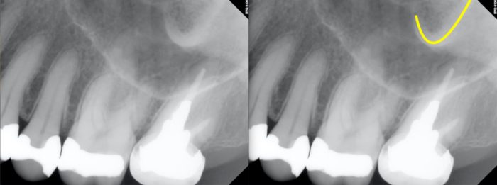
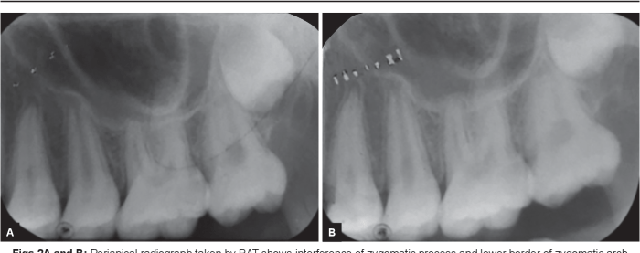
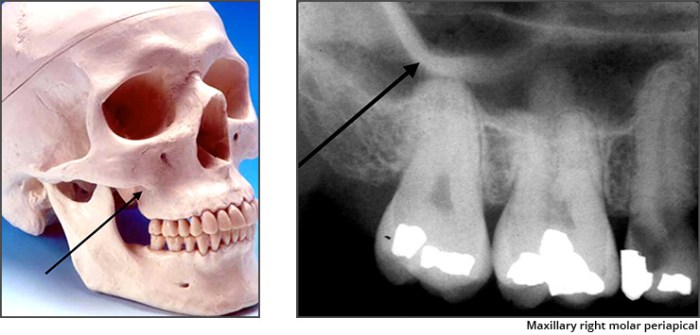
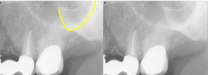
On panoramic radiographs, the zygomatic process appears as a radiopaque structure located superior to the maxillary sinus and lateral to the nasal cavity. On periapical radiographs, the zygomatic process may be seen as a radiopaque projection extending laterally from the maxilla.
On lateral cephalometric radiographs, the zygomatic process is visible as a radiopaque structure that forms the lateral wall of the orbit. The zygomatic arch can also be seen as a radiopaque line extending from the zygomatic process to the temporal bone.
Clinical Significance of Zygomatic Process Fractures
Zygomatic process fractures are relatively common facial injuries. They can be caused by blunt trauma to the face, such as a fall or a motor vehicle accident.
The clinical signs and symptoms of a zygomatic process fracture include pain, swelling, bruising, and difficulty opening the mouth. In some cases, the fracture may also cause diplopia (double vision).
Imaging Techniques for Zygomatic Process Assessment
CT and MRI are the imaging modalities of choice for evaluating zygomatic process injuries. CT provides excellent visualization of bony structures, while MRI can provide additional information about soft tissue injuries.
In cases of suspected zygomatic process fracture, a CT scan is typically the first imaging study performed. If there is concern about soft tissue injury, an MRI may also be ordered.
Treatment Options for Zygomatic Process Fractures
The treatment of zygomatic process fractures depends on the severity and location of the fracture. In some cases, conservative management with pain medication and ice packs may be sufficient.
In more severe cases, surgical intervention may be necessary to reduce the fracture and restore facial symmetry. Surgery may involve open reduction and internal fixation (ORIF) or closed reduction and internal fixation (CRIF).
FAQ Summary: Zygomatic Process Of Maxilla Radiograph
What is the zygomatic process of maxilla?
The zygomatic process is a projection of the maxilla that forms part of the lateral orbital rim and the infratemporal fossa.
How is the zygomatic process of maxilla visualized on radiographs?
The zygomatic process can be visualized on panoramic, periapical, and lateral cephalometric radiographs as a triangular or quadrilateral structure.
What are the clinical signs and symptoms of zygomatic process fractures?
Clinical signs and symptoms of zygomatic process fractures include facial swelling, ecchymosis, pain, and trismus.
What imaging techniques are used to assess zygomatic process injuries?
Imaging techniques used to assess zygomatic process injuries include CT and MRI.
What are the treatment options for zygomatic process fractures?
Treatment options for zygomatic process fractures range from conservative management to surgical intervention, depending on the severity and location of the fracture.


